Orbital Tumors Capillary Hemangioma
(Benign Hemangioendothelioma)
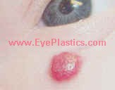
- A capillary hemangioma (sometimes referred to as an "Infantile hemangioma," "Strawberry hemangioma", and "Strawberry nevus") appears as a raised, red, lumpy area of flesh anywhere on the body, though 83% occur on the head or neck area. These marks occur in about 10% of all births, and usually appear between one and four weeks after birth. It may possibly grow rapidly, before stopping and slowly fading. Some are gone by the age of 2 , about 60% by 5 years, and 90–95% by 9 years.
- Capillary hemangiomas occur 5 times more often in female infants than in males, and mostly in Caucasian populations.] Additionally, low birthweight infants have a 26% chance of developing a hemangioma.
- It is the most common tumor of orbit and periorbital areas in childhood. While this birthmark may possibly be alarming in appearance, physicians generally counsel that it be left to disappear on its own, unless it is in the way of vision or blocking the nostrils.
-
General
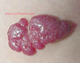
Color: strawberry or reddish-bluish-colored nevi
Texture: spongy
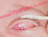
Location: upper eyelid more than lower eyelid; also occurs in the deep orbit (leads to proptosis)
Number: usually unilateral, often multiple
Family history: often
Size / Position: can increase in size with crying or positions in which they are lower to the ground
Presentation / Onset: appear in the initially eight l weeks of life
Rate of growth: rapidly for approximately six months to one year
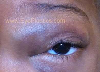
Natural history: spontaneous involution starts approximately at one year through age 6
Incidence: occurs in approximately 1-4% of infants, may possibly be more common in boys (3:2), more common in low birth weight infants.
- Systemic Evaluation
- occasional additional hemangiomas
- usually no other significant involvement (EXCEPTION: Kasabach-merritt Syndrome)
- Risks / Significance
- Vision can be affected in several ways
- inducement of strabismus which my lead to amblyopia
- occlusion of the visual axis
- inducement of astigmatism (related to position of tumor) and or myopia which may possibly lead to amblyopia; refractive error may possibly persist even after resolution of tumor
- Differential diagnosisImaging
- orbital cellulitis
- rhabodmyosarcoma
- lymphangioma
- orbital dermoid
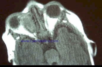
- CT - well circumscribed lesion
- MRI - well circumscribed lesion
Pathogenesis
Pathology
- tumor composed of numerous closely packed capillaries.
- proliferation of well-differentiated capillary endothelial cells
Treatment / Medical /Surgery
Not all orbital hemangiomas need to be removed. If, however, there is evidences of amblyopia or significant Ptosis ('Ptosis is sometimes referred to as Blepharoptosis. It refers to an eyelid which is droopy. This may possibly cause a loss of vision, especially while reading, headaches, and eyebrow strain.') treatment may possibly be initiated as outlined below:
- Amblyopia management
- Corticosterioid
- Oral
- Topical
- Local injection
- mixture of Kenalog and Celestone soluspan
- ObservationRadiation
- appropriate for small, non-visually threatening tumors
- Interferon
- CO 2 Laser